Lab One - Teamwork - Gram-Staining
LABORATORY REPORT
INTRODUCTION
In the Laboratory our primary goal was to identify the negative and positive gram-stained bacterium. Gram staining is a technique used to determine if a bacterium is gram -positive or gram -negative. In our lab we decided to gram stain a sample from the bacteria , E.Coli; yes the one that makes you sick, Staphylococcus epidermidis,and other samples obtained from random surfaces, well more like the inside of Debrina's nose and Carolann's tonsils with a swab. These samples were cultured for one week to inspect under the microscope and then gram-stained. The team believes we will find a selection of both gram-negative and gram-positive bacteria.
METHODS and MATERIALS
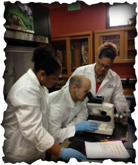
To start our procedure, using a bunsen burner, we sterilized a metal wire with a small loop at the end which will be used to handle the bacteria. We made sure the wire loop was steriled in the burning flame before and after each bacterial usage to guarantee proper bacterial smear and was cooled before obtaining the actual bacterial sample which we will refer to as the loop sterilizing process. We then placed the loop into deionized water after the sterilizng process and placed two small drops of water onto a microscopic slide.The metal loop was lit in the bunsen burner again (loop sterilizing process) until glowing red hot to sterilize and kill any bacteria which may be present. The metal loop was then cooled by touching the gel then inserted into the E.Coli cultured bacteria and stirred with the two drops of water to the size of a quarter on the slide. The loop steriling process was done again. With a wooden clip clasping the slide it was then passed through the bunsen flame a few times to warm the liquid into evaporate leaving a dry substance on the slide. The slide was not overheated to protect against cracking the slide and killing the bacteria. With the residue behind crystal- violet dye was added and let sit for a minute. The slide was then rinsed in three beakers of deionized water to clear the dye and then repeated with iodine solution for a minute then rinsed in the same fashion. Alcohol was then poured on the slide until the alcohol run clear before adding safranin-o, a red dye, for 30 seconds. The dye was rinsed three times before blotting with bibulous paper and then put under the mircroscope for inspection to verify gram negative or positive bacteria.
This process was repeated for S. epidermidis, throat swab, and tonsil swab.
RESULTS
The Team observed E. coli as a negative-gram stain rod-shaped bacterium under microscope. The Staphylococcus epidermidis was found to be a positive-gram stain bacterium in cohesive rounded colonies forming grape-like clusters. The team believed we would find positive and negative-gram bacterium but did not identify which bacterium was going to be negative or positive.
DISCUSSION AND ANALYSIS
This experiement demonstrated the two types of gram-staining bacterium and different types and shapes of bacterium. We also observed the bacterium S.epidermidis which was also a similar bacterium present on Carolann's tonsil swab and S.epidermidis is normally found in mucous membrances, are usually not a pathogenic bacterium but can be concerning for hospital patients.
Process of Gram-Staining Pictures
INTRODUCTION
In the Laboratory our primary goal was to identify the negative and positive gram-stained bacterium. Gram staining is a technique used to determine if a bacterium is gram -positive or gram -negative. In our lab we decided to gram stain a sample from the bacteria , E.Coli; yes the one that makes you sick, Staphylococcus epidermidis,and other samples obtained from random surfaces, well more like the inside of Debrina's nose and Carolann's tonsils with a swab. These samples were cultured for one week to inspect under the microscope and then gram-stained. The team believes we will find a selection of both gram-negative and gram-positive bacteria.
METHODS and MATERIALS
To start our procedure, using a bunsen burner, we sterilized a metal wire with a small loop at the end which will be used to handle the bacteria. We made sure the wire loop was steriled in the burning flame before and after each bacterial usage to guarantee proper bacterial smear and was cooled before obtaining the actual bacterial sample which we will refer to as the loop sterilizing process. We then placed the loop into deionized water after the sterilizng process and placed two small drops of water onto a microscopic slide.The metal loop was lit in the bunsen burner again (loop sterilizing process) until glowing red hot to sterilize and kill any bacteria which may be present. The metal loop was then cooled by touching the gel then inserted into the E.Coli cultured bacteria and stirred with the two drops of water to the size of a quarter on the slide. The loop steriling process was done again. With a wooden clip clasping the slide it was then passed through the bunsen flame a few times to warm the liquid into evaporate leaving a dry substance on the slide. The slide was not overheated to protect against cracking the slide and killing the bacteria. With the residue behind crystal- violet dye was added and let sit for a minute. The slide was then rinsed in three beakers of deionized water to clear the dye and then repeated with iodine solution for a minute then rinsed in the same fashion. Alcohol was then poured on the slide until the alcohol run clear before adding safranin-o, a red dye, for 30 seconds. The dye was rinsed three times before blotting with bibulous paper and then put under the mircroscope for inspection to verify gram negative or positive bacteria.
This process was repeated for S. epidermidis, throat swab, and tonsil swab.
RESULTS
The Team observed E. coli as a negative-gram stain rod-shaped bacterium under microscope. The Staphylococcus epidermidis was found to be a positive-gram stain bacterium in cohesive rounded colonies forming grape-like clusters. The team believed we would find positive and negative-gram bacterium but did not identify which bacterium was going to be negative or positive.
DISCUSSION AND ANALYSIS
This experiement demonstrated the two types of gram-staining bacterium and different types and shapes of bacterium. We also observed the bacterium S.epidermidis which was also a similar bacterium present on Carolann's tonsil swab and S.epidermidis is normally found in mucous membrances, are usually not a pathogenic bacterium but can be concerning for hospital patients.
Process of Gram-Staining Pictures
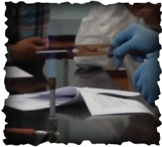
Evaporating the water from the bacteria sample over the bunsen burner flame
|
Gary washing the safranin-o dye from the bacteria slide
|
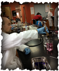
Results of gram-staining
Reference
Tacklyn, Barbara, C. "Swabs, Cultures, and Gram Staining". 2013. JPG file.
