Lab Two
CULTURE COMMON BACTERIA USING
ASEPTIC TECHNIQUE
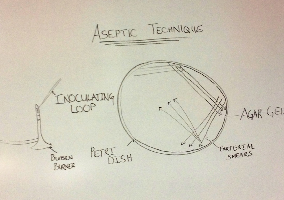
Sketch by Debrina Simons - March 2013
Introduction
In this Laboratory our primary goal was to learn the aseptic technique of culturing bacteria. Bacteria cultured were obtained from Carolann's tonsil and Debrina's nose. Bacteria that inhibit these areas are normally the S.epidermidis which are circular colonies in punctiform. S.epidermidis can be found on dirty surfaces such as an unclean keyboard or door knobs.
Material and Methods
A sterile swab and closed petri dish containing sterile nutrient agar was obtained. The sterile swab was than opened and inserted in Carolann's mouth. The tip of the swab was gently massaged across her tonsils. As a team we predicted that there would be many different colonies of bacterium growth because Carolann was having an painful irritation in her throat and because we know that there are many common bacteria that live on and in our bodies.
Next the perti dish was opened with very little exposure to the outer surrounding and the exposed swab was gently dragged across the agar in the same manner as shown in the above picture. The petri dish was than closed and taped shut with some parafin tape for seven days. The petri dish was turned upside and labeled with Carolann's initials, date and swab contents (Carolann's Tonsils). The dish was left facing down to avoid the water condensation droplets from falling onto the
agar and possibly contaminating the final results. On the next page in the picture slide show you will be able
to view the actual petri dish of the bacteria that grew from Carolann's tonsils over the seven day period of incubation.
Results
After a week of incubation the petri dishes showed remarkable growth of punciformed colonies under the light microscope. Although our team swabbed other surfaces we found Carolann's tonsils to be most fascinating. Her petri dish had grew many colonies. Theses colonies were whole colonies of punctiform, circular and irregular shapes. The colonies varied in size also. They ranged from punctiform to small, medium and large.
Discussion and Analysis
The Team after observing the results, discussed the possible bacterium found. The bacteria family Staphyloccaceae a known bacterium for the skin and human respiratory tract could potentially be the bacterial colonies observed but this can only be verified under farther observation (Wikipedia encyclopedia). When the swab was taken by Carolann she experiencing soreness in her throat. However over the cultures incubation period Carolann did not experience any further symptoms. But it was interesting to find that the results on the petri dish showed many growth colonies. This proves that there is bacteria that live on and in our bodies.
Reference
Tacklyn, Barbara, C. "Tonsil Swab". 2013. JPG file.
Wikipedia Encyclopedia, retrieved from website March 27, 2013, http://en.wikipedia.org/wiki/Staphylococcus_aureus
Wikipedia Encyclopedia, retrieved from website March 27, 2013, http://en.wikipedia.org/wiki/Staphylococcus_aureus
